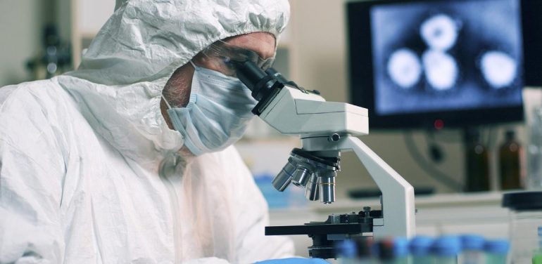
After 19 months of work and decades of previous research on coronaviruses, we now have a detailed description of how SARS-CoV-2 invades human cells. Scientists have discovered critical adaptations of the virus, which help it attach to human cells with astonishing power and then hide as soon as it invades them. Later, as it leaves the cells, SARS-CoV-2 performs a critical process to prepare the new virus particles to infect even more human cells. These are some of the tools that allowed the virus to spread so quickly - and why it is so difficult to control - and are reviewed in a publication in the journal Nature. The professors of the Therapeutic Clinic of the Medical School of the National and Kapodistrian University of Athens, Efstathios Kastritis, Theodora Psaltopoulou and Thanos Dimopoulos (rector of EKPA) present the main points of the publication.
The new SARS-CoV-2 coronavirus has a sugar coating, as shown in its computer simulations, and in particular in the pin proteins, which stand out on its surface. These sugar molecules are known as glucans.
Many viruses have glucans that mask their external proteins and camouflage them by hiding them from the immune system. Researchers, however, created a detailed representation of this cover based on structural and genetic data, which was eventually reconstructed person by person by a supercomputer, giving an extremely detailed representation. Within this cover, however, an uncoated loop stands out, which is a portion of the pin protein that constitutes the receptor binding domain (RBD), one of the three pin segments that bind to ACE2 receptors in human cells. .
In the simulation, when the RBD is projected over the sugar cover, two other glucan molecules take a position that locks the RBD in place, supporting it. However, the simulation models showed that if these glycans change then RBD collapses. Another research team developed a technique to test the same experiment in the laboratory, and by June 2020 they found that mutations in the two glycans reduced the ability of the spike protein to bind to the ACE2 receptor in human cells. This finding was not previously described in coronaviruses. Thus, it is possible that the rejection of these two sugars could reduce the infectivity of the virus, although researchers still do not have a way to do this.
From the beginning of the pandemic COVID-19, scientists have understood the details of how SARS-CoV-2 infects cells. By focusing on the infection process, they hope to find better ways to stop it through improved treatments and vaccines and learn why more recent strains, such as the Delta variant, are more contagious.
Each SARS-CoV-2 virus particle has an outer surface with 24-40 randomly arranged spike proteins that is the key to binding to human cells. For other types of viruses, such as influenza, the external binding / fusion proteins are relatively rigid. However, the pins of the SARS-CoV-2 are extremely flexible and articulate at three different points. This allows the pins to rotate and oscillate, making it easy to scan the cell surface and connect multiple peaks to a human cell at the same time. There are no similar experimental data for other coronaviruses, but because the spike protein sequences appear conserved in coronavirus evolution, it is probably a common feature of viruses in this family.
At the beginning of the pandemic, the researchers confirmed that the RBD region of the SARS-CoV-2 spike protein binds to the ACE2 receptor, which is located outside many cells of the upper respiratory tract and lung cells. This receptor is also the binding site of SARS-CoV, the virus that causes severe acute respiratory syndrome (SARS). However, compared to SARS-CoV, it is estimated that SARS-CoV-2 binds to ACE2 2-4 times more strongly because several changes in the RBD region stabilize the binding sites of the virus.
Of particular concern are SARS-CoV-2 variants that show mutations in the S1 subunit of the pin protein, which hosts the RBD region and is responsible for binding to the ACE2 receptor. (A second pin module, S2, helps fuse the virus envelope with the host cell membrane). The Alpha variant, for example, includes ten changes in the protein-spike sequence, which make RBD more likely to remain in a position that aids the virus, facilitating its entry into cells. The Delta variant, which is now rapidly spreading, hosts multiple mutations in the S1 subunit, including three in the RBD region that appear to enhance the RBD ability to bind to the ACE2 receptor and bypass the immune system.
Once the virus needles bind to the ACE2 receptor, other proteins on the surface of the host cell begin a process that leads to the fusion of the virus envelope and cell membrane.
The SARS-causing virus, SARS-CoV, uses one of the two protease enzymes (are enzymes that "cut" other proteins at specific sites) in the host cell to invade: TMPRSS2 or cathepsin L. TMPRSS2 is the fastest route, but SARS-CoV often enters through an endosome - a lipid-surrounded bubble - based on cathepsin L. However, when viruses enter cells in this way, antiviral proteins can trap them. .
SARS-CoV-2 differs from SARS-CoV in that it uses TMPRSS2 more efficiently, an enzyme found in high concentrations on the surface of respiratory epithelial cells. First, TMPRSS2 cleaves a site in the S2 subunit and thus exposes a series of hydrophobic amino acids that rapidly penetrate the nearest membrane: that of the host cell. The protruding pin is then folded over it, like a zipper, leading to the fusion of the virus envelope and the cell membrane.
The virus then launches its genome (its genetic material) directly into the cell. By invading in this way, like a spring, SARS-CoV-2 infects cells faster than SARS-CoV and avoids being trapped in endosomes.
The rapid entry of the virus, using TMPRSS2, explains why chloroquine did not work in clinical trials as a treatment for COVID-19, despite the first promising laboratory studies. Laboratory studies used cells that used only cathepsin to enter the virus through endosomes. When the virus is transmitted and reproduces in the human respiratory epithelium it does not use endosomes, so chloroquine, which is a drug that inhibits endosomes, is not effective.
This finding also suggests that protease inhibitors could be a promising treatment that would inhibit the virus' ability to use TMPRSS2, cathepsin L, or other proteases to enter cells. An inhibitor of TMPRSS2, mesylate kamostate, which has been approved in Japan for the treatment of pancreatitis, prevented the virus from entering the lung cells, but the drug did not improve patients' outcome in an initial clinical trial.
The next stages of the infection are less clear. After the virus injects its genome (which is in the form of RNA) into the cell, then the ribosomes in the cytoplasm translate two segments of the virus RNA into large amino acid sequences, which are then broken down into 16 proteins, including many involved in its synthesis. RNA. Later, more RNA molecules are generated encoding a total of 26 known viral proteins, including structural proteins used to produce new virus particles, such as spike and other helper proteins. In this way, the virus begins to make copies of its own messenger RNA. But it needs the cellular mechanism to translate these mRNAs into proteins. Coronaviruses control this mechanism in many ways. Researchers have focused on three mechanisms by which SARS-CoV-2 suppresses host mRNA translation in favor of its own. Although no one is unique to this virus, the combination, speed and effectiveness seem unique. First, the virus eliminates competition: The Nsp1 virus protein, one of the first proteins to be translated when the virus arrives, recruits host proteins that systematically cut all mRNA molecules into the cell that are not labeled as viral. When researchers put a similar label on a host cell mRNA molecule then the mRNA is not cut into pieces.
Second, infection reduces the total protein translation in the cell by 70%. The Nsp1 virus protein is again the main culprit, this time blocking the ribosome entry channel so that mRNA cannot enter and the remaining limited translation capacity is dedicated to the virus mRNA molecules.
Finally, the virus "closes" the cell alarm system. This happens in many ways, but basically the virus prevents cellular mRNA from coming out of the nucleus, including instructions for proteins intended to alert the immune system to infection. Again, Nsp1 appears to block output channels in the cell nucleus. Because gene transcripts in mRNA cannot leave the nucleus, infected cells do not release much interferon, which normally alerts the immune system to the presence of a virus. SARS-Cov-2 is particularly effective in inhibiting this alarm system: Compared to other respiratory viruses, including SARS-CoV and respiratory syncytial virus, SARS-CoV-2 infection causes significantly lower levels of interferons. In addition, researchers report that mutations in the alpha variant appear to make it more capable of inhibiting interferon production even more effectively.
Once the virus takes control of the translation mechanism, it extensively reshapes the inside and outside of the cell according to its needs.
First, some of the newly assembled virus-spike proteins travel to the cell surface and push the host membrane. There, they activate a channel of calcium ions in the host, which eliminates a fatty coating on the outside of the cell, the same coating found on fused cells as muscle cells. At this point, the infected cells fuse with neighboring cells expressing the ACE2 receptor and develop into bulky respiratory cells filled with up to 20 nuclei. These fused structures, called syncytia, are caused by other viral infections, such as HIV and herpes simplex, but not by SARS. Possibly the formation of syncytia allows the infected cells to thrive for long periods of time, giving rise to more and more virus. Some infected cells form clots even with lymphocytes, one of the cells of the immune system. This is a known mechanism for protecting the immune system from cancer cells, but not from viruses.
Even greater changes occur inside the cell. Like other coronaviruses, SARS-CoV-2 transforms the long, thin endoplasmic reticulum (ER), a network of flat membranes involved in protein synthesis and transport, into double-membrane spheres, as if the endoplasmic reticulum forms bubbles. These dual membrane vesicles (DMVs) may provide a safe place for viral RNA to reproduce and translate, protecting it from the cell's innate immune sensors, but this hypothesis is still being investigated.
The proteins involved in the formation of double-membrane vesicles could be good targets for drugs because they appear to be essential for virus replication. For example, a host cell protein, TMEM41B, is required to mobilize cholesterol and other lipids to extend the membranes of the endoplasmic reticulum so that all parts of the virus fit inside. Inhibition of TMEM41B has a significant impact on infection. Coronavirus Nsp3 transmembrane protein could also be a target, as it creates a crown-like pore in the walls of double-membrane vesicles to remove viral RNA produced after infection.
Most viruses that have an outer sheath, known as an envelope, form this feature by assembling the vesicles directly on the edge of the cell, stealing portions from the cytoplasmic membrane of the cell itself as they exit. But newly formed coronavirus proteins follow a different path.
Evidence shows that coronaviruses are transported out of the cell through the Golgi complex, which is an organ that functions as a mail: It packs molecules into membranes and sends them to other parts of the cell. There, the virus forms a lipid envelope from the membrane of the Golgi complex. The newly formed virus particles are then transported into the Golgi vesicles to the cell surface, from where they are ejected out of the cell.
But researchers in the United States say they have detected coronaviruses leaving the cell through lysosomes (they are intracellular "garbage" bins filled with enzymes that break down parts of cells). They observed that blocking the secretory pathway based on the Golgi system did not appear to affect the amount of infectious virus released. In contrast, the evidence suggests that the virus proteins form an envelope within the endoplasmic reticulum and then exploit the lysosomes to leave the cell. Researchers are currently testing inhibitors that block the lysosomal exit process.
Leaving a cell through the Golgi system or lysosomes is slow and inefficient compared to hatching from the cytoplasmic membrane, and scientists do not know why SARS-CoV-2 does this. It is suspected that the lipid synthesis of an envelope derived from Golgi or lysosomes is perhaps more "suitable" for the virus than from an envelope derived from the cytoplasmic membrane.
Upon leaving the cell, another fact makes this virus highly contagious: A rapid cleavage at a site with five amino acids prepares the virus to achieve its next goal. Where other coronaviruses have only one amino acid arginine at the junction of the S1 and S2 subunits of the spike, SARS-CoV-2 has a sequence of five amino acids. In May 2020, researchers reported that a host cell protein called furin recognizes and cuts down this amino acid sequence, and this cut is necessary for the virus to enter the human lung cells efficiently.
This is not the first time researchers have identified a cleavage site for furine in a virus. The highly pathogenic avian influenza viruses also have such sites. A strain of SARS-CoV-2 in culture lost this cleavage site from furine and showed a lower ability to infect cells in experimental animals. Another study also found that coronavirus with an intact furin cleavage site enters human airway cells faster than without this cleavage site.
The furine probably "cuts" this area at some point during the assembly of the virus particles or shortly before their release. The synchronization may explain why the virus exits through the Golgi or lysosomes, as once the virus is assembled it is transported to an organelle where the protease furin is abundant.
By detaching the "link" between subunits S1 and S2, after cleavage by furin, the spike of the virus will undergo a second "cut" by TMPRSS2, which exposes the hydrophobic region that is rapidly "buried" in the cell membrane. host. If the spikes are not pre-treated by the furin - and this is not always the case - then they are forced to skip the treatment stage by TMPRSS2 and enter through the slower path through the bays or not at all.
Alpha and Delta mutations
Two variants of the coronavirus, Alpha and Delta, have altered furin cleavage sites. In the Alpha variant, the original amino acid proline is changed to histidine (P681H), while in the Delta variant it is changed to arginine (P681R). Both changes make the sequence less acidic and the more basic the amino acid sequence, the more efficiently furin recognizes and cuts it. As a result, the virus becomes even more efficient in its transmission. More spikes "cut" by furin means more spikes entering human cells. In SARS-CoV, less than 10% of spike proteins are "processed", but in SARS-CoV-2 this percentage increases to 50%. In the Alpha variant, it exceeds 50%. In the highly contagious Delta variant, over 75% of the spikes are prepared to infect a human cell.
There are still many unknown factors to understand COVID-19. Key knowledge gaps include the number of ACE2 receptors required to bind to each spike-protein, when the S2 subunit cleaves by TMPRSS2, and the number of spikes required to fuse between viruses and cell membranes. Meanwhile, at least 332 interactions between SARS-CoV-2 and human proteins have been identified. Most mutations, so far, are related to how effectively the virus spreads, not how much the virus destroys the host, experts agree. The Delta variant is characterized by a faster increase in virus levels and higher levels in the airway than previous variants of the virus, but it is not yet fully understood how the Delta mutations have made the virus so contagious.
Source: RES-EAP
